Full Mouth X-ray
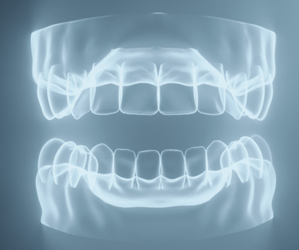
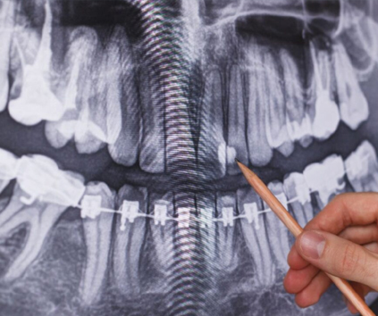
Dental braces are orthodontic devices designed to correct misalignments and irregularities in the positioning of teeth. This widely used orthodontic treatment helps improve both the function and appearance of the teeth and jaws. Here are key aspects of dental braces:
Types of Dental Braces:
Traditional Braces: These consist of metal brackets attached to each tooth, connected by wires and secured with rubber bands. They are highly effective and have been used for many years.
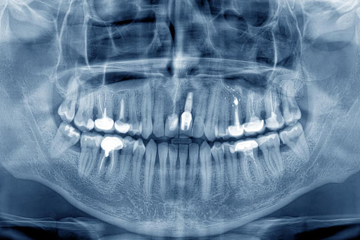
A full mouth X-ray, also known as a panoramic radiograph or orthopantomogram (OPG), is a diagnostic imaging technique used in dentistry to capture a comprehensive view of the entire oral cavity, including the teeth, jaws, and surrounding structures. This non-invasive and efficient imaging method offers numerous benefits in dental diagnostics and treatment planning.
Comprehensive Visual of Oral Structures:
A full mouth X-ray provides a detailed and panoramic view of the upper and lower jaws, all the teeth, temporomandibular joints (TMJ), and adjacent structures. This allows dentists to assess the overall condition of the oral cavity in a single image.
Diagnostic Capabilities:
Dentists use full mouth X-rays to diagnose a wide range of dental issues, including cavities, gum diseases, impacted teeth, cysts, tumors, and abnormalities in the jawbone. It is a valuable tool for detecting conditions that may not be visible during a routine clinical examination.
Treatment Planning:
The information obtained from a full mouth X-ray is crucial for treatment planning in various dental procedures. Orthodontists use it to assess the alignment of teeth, oral surgeons use it to plan extractions or implant placements, and general dentists use it to formulate comprehensive treatment plans.
Convenience and Patient Comfort:
Obtaining a full mouth X-ray is a relatively quick and comfortable procedure for the patient. The imaging equipment rotates around the head, capturing a complete image without the need for intraoral films or uncomfortable positioning.
Identification of Impacted Teeth:
Full mouth X-rays are particularly useful in identifying impacted teeth, such as wisdom teeth, and assessing their position and potential impact on adjacent structures.
Bone Health Assessment:
The X-ray provides valuable information about the density and health of the jawbone. This is crucial for assessing the viability of certain dental procedures, such as dental implant placement.
Radiation Safety Measures:
Modern dental X-ray technology employs low levels of radiation, and additional safety measures are often implemented, such as lead aprons, to minimize radiation exposure to other parts of the body.
Integration with Other Diagnostic Tools:
Full mouth X-rays complement other diagnostic tools, including intraoral X-rays and clinical examinations. The combination of these methods provides a comprehensive understanding of a patient’s oral health.
Digital Advancements:
Many dental practices have transitioned to digital full mouth X-rays, which offer advantages such as reduced radiation exposure, immediate image availability, and enhanced image quality. Digital images can also be easily stored and shared electronically.
In conclusion, a full mouth X-ray is a valuable diagnostic tool that plays a central role in comprehensive dental care. It aids in early detection, diagnosis, and treatment planning, contributing to effective and personalized oral health management. Regular use of full mouth X-rays is determined based on individual patient needs and specific dental conditions.